


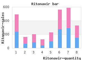
Almudena Burillo, M.D., Ph.D.
- Physician
- Clinical Microbiology and Infectious Diseases
- Hospital de Mostoles
- Madrid, Spain
The role of acid reduction by H blockade or with2 proton pump inhibitors to reduce the risk of invasive malignancy when these precursor lesions are found is unclear in treatment online buy on line ritonavir. Operations for benign peptic ulcers also appear to be associated with an increased risk of gastric cancer symptoms 9dp5dt buy ritonavir 250mg on-line. Adenocarcinoma in the gastric remnant may appear several decades after the index peptic ulcer operation and appears to increase over time treatment questionnaire purchase 250mg ritonavir amex. Finally medications kidney failure ritonavir 250 mg with visa, chronic medicine bg discount 250mg ritonavir, nonhealing gastric ulcers must be carefully evaluated endoscopically with multiple biopsies to determine if an underlying invasive malignant component is present medicine 4212 buy ritonavir line. All of these risk factors contribute a small marginal increase to the lifetime risk of gastric cancer. Even in the presence of these risk factors, the absolute lifetime risk may still be small. Genetics Most gastric carcinomas occur sporadically, however, 1% to 3% of them have an inherited familial component. Endoscopic surveillance is not a reliable strategy to prevent invasive malignancy in these patients. In general, the operation is performed when the patient is young (in their 20s), or no later than 10 years prior to the earliest age of gastric cancer diagnosed in their family. Virtually all carriers of this mutation have 382 multiple foci of intramucosal adenocarcinoma in the resected specimen. Gastric tumors are classified according to their site in the proximal (cardia) and distal (noncardia) stomach. Although the incidence of distal gastric cancer is decreasing in the United States, the incidence of proximal gastric tumors continues to increase. Cancers of the gastric cardia currently account for nearly 50% of all cases of gastric adenocarcinoma. Gastric cardia tumors are five times more common in men than women, whereas noncardia gastric tumors are twice as common in men as in women. In general, gastric tumors are more common on the lesser curve of the stomach than on the greater curve. The most common classification scheme is a two-category system called the Lauren classification. According to the Lauren classification, there are two histologic types of gastric adenocarcinoma: intestinal and diffuse. The intestinal type is found in geographic regions where there is a high incidence of gastric cancer and is characterized pathologically by the tendency of malignant cells to form glands. These tumors are usually well to moderately differentiated and associated with metaplasia or chronic gastritis. These tumors tend to spread through the lymphatics and hematologically to distant organs, most often the liver. The diffuse type typically lacks organized gland formation, is usually poorly differentiated, and has many signet ring cells. If more than 50% of the tumor contains intracytoplasmic mucin, then it is designated as signet ring type. Diffuse-type tumors are more common in younger patients with no history of gastritis and spread transmurally and by lymphatic invasion. Although the incidence of these tumors varies 383 little from country to country, their overall incidence appears to be increasing worldwide. The Lauren classification has its limitations, which include difficulty in assessing the classification on limited endoscopic biopsies and inability to classify patients who have had a significant response to preoperative therapy. An alternative classification scheme is used by the World Health Organization, which classifies gastric adenocarcinomas according to their histologic appearance, which includes tubular, mucinous, papillary, and signet ring cell types. Patients and physicians often ignore the vague epigastric discomfort that in hindsight was present for months prior to diagnosis. The most frequent presenting symptoms are weight loss, pain, vomiting, and anorexia. Tumors in the antrum may cause symptoms of gastric outlet obstruction; this is an ominous sign of advanced disease. Although rare, large tumors that directly invade the transverse colon may present with colonic obstruction. Chronic anemia is also a common finding that prompts endoscopic evaluation that reveals an otherwise occult gastric cancer. Physical examination findings that suggest metastatic disease include a palpable supraclavicular lymph node (Virchow node), a mass palpable on rectal examination (Blumer shelf), a palpable periumbilical mass (Sister Mary Joseph node), or the presence of ascites or jaundice. Intestinal-type gastric cancer most commonly metastasizes to the liver, while peritoneal metastases are the most common site of metastases for diffuse type. The recommended initial evaluation includes complete history and physical examination and 384 laboratory studies. Nutrition labs may be helpful to identify malnutrition and to guide decision making regarding operative timing. This allows early identification of solid organ metastases, malignant ascites, or possibly large peritoneal metastases. Esophagogastroduodenoscopy allows for direct anatomic localization of the tumor and sampling of tissue for diagnostic purposes. Staging laparoscopy is also a critical part of the initial assessment and staging of patients with newly diagnosed gastric adenocarcinoma. The intent is to identify metastatic peritoneal disease, therefore nodal biopsy is not routinely performed. Patients with microscopically positive peritoneal cytology findings have a prognosis similar to that of patients with macroscopic visceral or peritoneal disease. A repeat diagnostic laparoscopy prior to planned resection in patients with locally advanced disease after neoadjuvant treatment can also be considered and may identify radiographically occult metastatic disease in a minority of patients. The Japanese classification historically refers to the extent of lymph node dissection by D classification. The staging system relies on the classic tumor (T), node (N), and metastasis (M) classification. The updated eighth edition defines clinical, pathologic, and postneoadjuvant therapy stage groupings. Stages are grouped to identify survival differences, thus there is a mix of T and N classifications in each stage designation (Table 9. When comparing studies from different time periods, care must be paid 386 to changes in staging definitions. The T classification regarding depth of invasion was aligned with other gastrointestinal tract malignancies, such as the esophagus and small and large bowel. The modification of T stage in the seventh edition from previous editions include: tumor penetrates subserosal tissue (previously T2b, currently T3), serosa (previously T3, currently T4a), and adjacent structures (previously T4, currently T4b). The evaluation of the lymph nodes and the extent of lymph node resection have always been contentious issues in gastric cancer. A pN0 designation can be assigned based on the actual number of lymph nodes evaluated microscopically. The presence of metastatic disease is simply designated M1 without subclassification. Metastases to nonregional lymph node groups, including the retropancreatic, para-aortic, portal, retroperitoneal, or mesenteric groups, is considered M1 disease. Microscopically positive peritoneal cytology, in addition to frankly macroscopic peritoneal disease, is considered M1 disease. Japanese Classification Since 1962 when the first version of the Japanese Classification of Gastric Cancer was published, frequent modifications have been applied up to the current 14th version. The Japanese Classification not only defines 390 pathologic staging, but also describes all aspects of preoperative staging, intraoperative findings, how to handle resected specimens, rules of treatment effect on pathology, and detailed histologic classifications. There are also a series of Japanese Treatment Guidelines of Gastric Cancer, which summarize current consensus of stage-specific treatment strategies; currently it is on its fourth edition. The greatest advantage of the detailed Japanese classification is the standardization of the description of specific tumor status, which creates a standard format used by Japanese surgeons in each case. The disadvantages include its complexity which limits its broader acceptance and the frequent updates which make it difficult to compare results to historical literature. The current interest in Japan is to create a new classification for Siewert type 2 tumors, and a prospective study is underway to define the optimal surgical approach (right thoracotomy vs. The type of surgery is then dependent upon tumor depth, growth pattern, and tumor location. All other surgical resections are based on the location of the tumor and potential lymph node metastases, with the intent of microscopically negative surgical margins and clearance of the at risk lymph node groups. Endoscopic Mucosal Resection Gastric adenocarcinoma confined to the mucosa (T1a lesions) has a very 391 low rate of lymph node metastases, on the order of 0% to 5%. As such, if complete resection of these lesions is possibly via an endoscopic approach, acceptable locoregional disease control can be achieved with minimal comorbidities. The expanded indications include nonulcerated, well-differentiated lesions greater than 2 cm in diameter; ulcerated, well-differentiated lesions less than 2 cm in diameter; and undifferentiated, nonulcerated T1a lesions less than 2 cm in diameter. However, reports have described the use of expanded criteria such as resection of larger tumors, tumors with submucosal invasion, and for patients whose comorbidities preclude standard treatment with gastrectomy. The reported experience is mostly from eastern Asia, where intense surveillance programs are in place and the incidence of early T1a gastric cancer is high. As such, experience with this complicated endoscopic technique is very high in these countries. Gastric cancer is rarely diagnosed at such an early stage in Western countries lacking dedicated surveillance programs. The approach remains an option for treatment in very early-stage gastric cancer, but the appropriate endoscopic expertise and commitment to long-term follow-up are necessary. The approach can be abdominal, abdominothoracic, transhiatal, or transthoracic, via either an open or minimally invasive technique. This is performed with input from both the thoracic and surgical oncology teams, usually via an open transabdominal approach. We favor this approach so that a D2 lymph node dissection including the greater curvature lymph nodes can be performed in these patients. We do not in general perform proximal subtotal gastrectomies for these lesions in order to avoid alkaline reflux esophagitis. Tumors located in the body and antrum make up the remaining 65% to 70% of gastric cancers. The decision to make is whether to perform a distal, subtotal, or total gastrectomy in these patients. As long as a margin negative resection is performed, survival is equivalent among these operations. Quality of life and long-term nutrition status appears to be better in subtotal versus total gastrectomy. In these patients, we favor whatever extent of resection is needed to achieve a gross 5 cm margin. An adequate remnant proximal gastric pouch needs to be present to consider a subtotal gastrectomy. We do not perform a proximal subtotal gastrectomy with an esophagogastrostomy in more proximal body tumors, as mentioned above. Lymphadenectomy the extent of lymphadenectomy has been a much debated topic in gastric cancer treatment over the last several decades. Japanese surgeons have espoused radical lymphadenectomy and meticulous pathologic analysis, 393 with low perioperative morbidity and mortality. Western studies attempting to replicate these findings initially demonstrated alarmingly high rates of perioperative morbidity and mortality without survival benefit in extended lymphadenectomy. More recent data have supported more extensive lymphadenectomy for advanced disease, as long as it can be performed by experience surgeons with good outcomes. The Japanese definition of D1 or D2 lymph node dissection is complicated even after recent simplification. The extent of D1/D2 lymph node dissection changes based on the extent of gastrectomy, and D3 is no longer defined. An important update of the D1/D2 classification in the current Japanese classification is that the left gastric artery lymph nodes (#7) should be included in a D1 lymph node dissection in addition to perigastric lymph nodes, regardless of the procedure type or tumor location. The right paracardial lymph nodes (#1) are also included in a D1 lymph node dissection in any tumor location or procedure type, whereas the left paracardial lymph nodes (#2) and splenic hilum nodes (#10) are not included in either a D1 or D2 dissection for distal tumors. These lymph nodes should not be dissected in antral or distal body tumors that can be resected with distal gastrectomy. A lymph node dissection is classified as D0, D1, or D2; D0 refers to an incomplete resection of N1, D1 denotes a complete resection of N1, and D2 involves a complete removal of N1 and N2 lymph node stations. The extended D3 lymph node dissection, while it is no longer defined as such, includes further extent of lymph nodes such as para-aortic (#16) and retropancreatic (#13) lymph nodes. Longer-term follow-up in the Dutch trial reported a statistically significant difference in overall survival, favoring D2 (15-year survival 29% vs. The Italian study, with strict quality control measures regarding definition of D1 and D2 dissections and the experience of the participating surgeons, reported much lower perioperative morbidity and mortality rates, reassuring the medical community that experienced surgeons could perform extended lymphadenectomy for gastric cancer safely. The Japanese Clinical Oncology Group Study 9501 reported excellent perioperative outcomes for D2 and D3 lymphadenectomies, without long-term survival benefit from the addition of a D3 dissection of the para-aortic lymph nodes. A limited D1 lymphadenectomy may be sufficient for early stage gastric cancer, particularly in low-volume centers that may have higher complication rates.
Manipulate the abdominal hand gently downward toward the vaginal fingers to outline the adnexa medicine klimt discount ritonavir 250 mg with mastercard. A normal ovary (about 4 fi 2 fi 3 cm in size medications vertigo ritonavir 250 mg sale, sensitive medications contraindicated in pregnancy 250 mg ritonavir sale, firm treatment modalities 250 mg ritonavir amex, and freely movable) is often not palpable 97110 treatment code discount ritonavir master card. If an adnexal mass is found medicine mound texas buy 250mg ritonavir with mastercard, evaluate its location relative to the uterus and cervix, architecture, consistency, tenderness, and mobility. Palpate the left adnexal region, repeating the technique described previously, but place the vaginal fingers in the left fornix and the abdominal hand on the left lower quadrant. Insert the index finger into the vagina and the middle finger into the rectum very gently. The use of this technique makes possible higher exploration of the pelvis because the cul-de-sac does not limit the depth of the examining finger. Rectal examination Inspect the perianal and anal area, the pilonidal (sacrococcygeal) region, and the perineum for the following aspects: Color of the region (note that the perianal skin is more pigmented than the surrounding skin of the buttocks and is frequently thrown into radiating folds) Lesions 1. Pruritus ani is usually indicated by thickening, excoriations, and eczema of the perianal region and adjacent areas. The anal opening often is the site of fissures, fistulae, and external hemorrhoids. Palpate the pilonidal area, the ischiorectal fossa, the perineum, and the perianal region before inserting the gloved finger into the anal canal. Note the presence of any concealed induration or tenderness in any of these areas. Lay the pulp of the index finger against the anal orifice and instruct the subject to strain downward. Then, with a slight rotary movement, insinuate the finger past the anal canal into the rectum. Evaluate the anal canal for: Tonus of the external sphincter muscle and the anorectal ring at the anorectal junction Tenderness (usually caused by a tight sphincter, anal fissure, or painful hemorrhoids) Tumor or irregularities, especially at the pectinate line Superior aspect: Reach as far as you can. Mild straining by the patient may cause some lesions, which are out of reach of the finger, to descend sufficiently low to be detected by palpation. Test for occult blood: Examine the finger after it is withdrawn for evidence of gross blood, pus, or other alterations in color or consistency. Cervix: size, shape, symmetry, consistency, and tenderness, especially on manipulation 2. Rectouterine fossa for tenderness or implants In patients with an intact hymen, the examination of the anterior wall of the rectum is the usual method of examining the pelvic organs. Pelvic Examination the pelvic examination is usually performed with the patient in the dorsal lithotomy position (Fig. Raising the head of the examination table, if possible, may facilitate relaxation. Evidence of any lesions, erythema, pigmentation, masses, or irregularity should be noted. The skin quality should be noted as well as any signs of trauma, such as excoriations or ecchymosis. Areas of erythema or tenderness are noted, particularly in women with vulvar burning or pain, as might be seen with vulvar vestibulitis or localized provoked vulvodynia. The presence of any visible lesions should be quantitated and carefully described with regard to their full appearance and characteristics on palpation. Ulcerative or purulent lesions of the vulva should be evaluated and cultured as outlined in subsequent chapters, and biopsy should be performed on any lesions. It may be helpful to ask the patient if she is aware of any vulvar lesions and to offer a mirror to demonstrate any lesions. After thorough visualization and palpation of the external genitalia, including the mons pubis and the perianal area, a speculum is inserted into the vagina. The smallest width speculum necessary to produce adequate visualization should be used. The larger Graves speculum may be required in women who have lax vaginal walls, are pregnant, or will be undergoing cervical or endometrial biopsies or procedures. In some women, a longer speculum (either Pederson or Graves) may facilitate visualization of the cervix in a manner that is less uncomfortable to the patient. If any speculum other than the typically sized specula is used, the patient should be informed and encouraged to remind the clinician before her next examination. The speculum should be warmed before it is inserted into the vagina; a heating pad or speculum warmer should be placed under the supply of specula. If lubrication is required, warm water generally is sufficient or a small amount of lubricant can be used without interfering with cervical cytology testing. The patient should be asked to relax the muscles of her distal vagina before the insertion of the speculum to facilitate the placement and to avoid startling her by this portion of the examination. After insertion, the cervix and all aspects of the vagina should be carefully inspected. The appropriate technique and frequency for cervical cytology testing is presented in Chapter 19. An endometrial biopsy usually is performed with a flexible cannula or a Novak curette (see Chapter 14). Testing for sexually transmitted diseases should be performed routinely in adolescents and young adults as recommended by the Centers for Disease Control and Prevention. After the speculum is removed and the pelvis palpated, lubrication is applied to the examination glove, and one or two (the index or index and middle) fingers are inserted gently into the vagina. In general, in right-handed physicians, the fingers from the right hand are inserted into the vagina and the left hand is placed on the abdomen to provide counter-pressure as the pelvic viscera are moved (Fig. The vagina, its fornices, and the cervix are palpated carefully for any masses or irregularities. One or two fingers are placed gently into the posterior fornix so the uterus can be moved. With the abdominal hand in place, the uterus usually can be palpated just above the surface pubis. In this manner, the size, shape, mobility, contour, consistency, and position of the uterus are determined. The patient is asked to provide feedback about any areas of tenderness, and her facial expressions are observed during the examination. The adnexa are palpated gently on both sides, paying particular attention to any enlargements. Again, the size, shape, mobility, and consistency of any adnexal structures should be carefully noted. When indicated, a rectovaginal examination should be performed to evaluate the rectovaginal septum, the posterior uterine surface, the adnexal structures, the uterosacral ligaments, and the posterior cul-de-sac. Uterosacral nodularity or posterior uterine tenderness associated with pelvic endometriosis or cul-de-sac implants of ovarian cancer can be assessed in this manner. Hemorrhoids, anal fissures, sphincter tone, rectal polyps, or carcinoma may be detected. A single stool sample for fecal occult blood testing obtained in this manner is not adequate for the detection of colorectal cancer and is not recommended (55) (Fig. At the completion of the physical examination, the patient should be informed of the findings. When there is a possible abnormality, the patient should be informed immediately; this discussion should take place after the examination with the patient clothed. A plan to evaluate the findings should be outlined briefly and in clear, understandable language. Pediatric Patients A careful examination is indicated when a child presents with genital symptoms such as itching, discharge, burning with urination, or bleeding. The examiner should be familiar with the normal appearance of the prepubertal genitalia. The normal unestrogenized hymenal ring and vestibule can appear mildly erythematous. The technique of examination is different from that used for examining an adult and may need to be tailored to the individual child based on her age, size, and comfort with the examiner. A speculum examination should not be performed in a prepubertal child in the office. The child who is relaxed and warned about touching will usually tolerate the examination satisfactorily. Two percent lidocaine jelly may be used as a topical anesthetic to facilitate the examination if needed. Some children who were abused, who had particularly traumatic previous examinations, or who are unable to allow an examination may need to be examined under anesthesia, although a gentle office examination should almost always be attempted first. If the child had bleeding and no obvious cause of bleeding is visible externally or within the distal vagina, an examination under anesthesia is indicated to visualize the vagina and cervix completely. A hysteroscope, cytoscope, or other endoscopic instrument can be used to provide magnification and as a light source for vaginoscopy, which should be performed under anesthesia. An adolescent who presents with excessive bleeding should have a pelvic examination if she had intercourse, if the results of a pregnancy test are positive, if she has abdominal pain, if she is markedly anemic, or if she is bleeding heavily enough to compromise hemodynamic stability. The pelvic examination occasionally may be deferred in young teenagers who have a classic history of irregular cycles soon after menarche, who have normal hematocrit levels, who deny sexual activity, and who will reliably return for follow-up. Current guidelines recommend that cervical cytology testing in most adolescents be initiated at age 21 (58). Other diagnostic techniques (such as pelvic ultrasound) can substitute for or supplement an inadequate examination. An examination usually is required when there is a question of pelvic pain, genital anomaly, pregnancy-related condition, or possibility of pelvic infection. It is helpful to ascertain whether the patient had a previous pelvic examination, how she perceived the experience, and what she heard about a pelvic examination from her mother or friends. Before a first pelvic examination is performed, a brief explanation of the planned examination (which may or may not need to include a speculum), instruction in relaxation techniques, and the use of lidocaine jelly as a lubricant can be helpful. The patient should be encouraged to participate in the examination by voluntary relaxation of the introital muscles or by using a mirror if she wishes. If significant trauma is suspected or the patient finds the examination too painful and is truly unable to cooperate, an examination under anesthesia may be necessary. The risks of general anesthesia must be weighed against the value of information that would be obtained by the examination. Particularly with regard to issues as sensitive as sexual activity, it is critical that the adolescent be interviewed alone, without a parent in the room. The patient should be asked whether she engaged in sexual intercourse, whether she used any method of contraception, used condoms to minimize the risks of sexually transmitted diseases, or she feels there is any possibility of pregnancy. Follow-Up Arrangements should be made for the ongoing care of patients, regardless of their health status. Patients with no evidence of disease should be counseled regarding health behaviors and the need for routine care. For those with signs and symptoms of a medical disorder, further assessments and a treatment plan should be discussed. The physician must determine whether she or he is equipped to treat a particular problem or whether the patient should be directed to another health professional, either in obstetrics and gynecology or another specialty, and how that care should be coordinated. If the physician believes it is necessary to refer the patient elsewhere for care, the patient should be reassured that this measure is being undertaken in her best interests and that continuity of care will be ensured. Patients deserve a summary of the findings of the visit, recommendations for preventive care and screening, an opportunity to ask any additional questions, and a recommendation for the frequency of any follow-up or ongoing care visits. Physicians are challenged to practice the art of medicine in a manner that leads to effective alliances with their patients. Physicians should listen carefully to what patients are saying about the nature and severity of their symptoms. They should listen to what patients may not be expressing: their fears, anxieties, and personal experiences that lead them to react in a certain manner when faced with what is often, to them, a crisis (such as the diagnosis of an abnormality on examination, laboratory testing, or pelvic imaging). Physicians should supplement their formal education and clinical experience by constantly seeking valid new information and honing their communication skills. To meet the challenges posed by the complexities of patient care, physicians must learn to practice evidence-based medicine, derived from the very latest data of highest quality.
If there are more than 200 individual case reports treatment west nile virus order ritonavir 250mg on line, submit only summary tabulations and not line-listings treatment 2015 buy ritonavir 250 mg cheap. If subsequently requested by a regulator treatments yeast infections pregnant purchase ritonavir 250 mg online, however medicine 0025-7974 buy generic ritonavir on line, a line listing should be provided within 10 working days treatment scabies buy cheap ritonavir 250mg line. For a five year gap between reports treatment of diabetes discount 250 mg ritonavir otc, follow-up information on cases described in the previous report should only be provided for cases associated with ongoing or new safety issues. Inclusion and discussion of literature reports should be selective and focus on publications relevant to safety findings, independent of listedness. For reports with extensive numbers of case reports, discussion and analysis for the Overall Safety Evaluation should be partitioned by system organ class, rather than by listedness or seriousness. In general, the criteria that should be considered for an abbreviated report are: no serious unlisted cases and few. In an abbreviated report, it should be unnecessary to include the usual full inventory of locations where the drug is marketed. Proposals Relating to Frequency and Timing of Reporting A Summary Bridging Report. It will ordinarily supplement annual or five year reports and should not be required routinely but only on special regulatory request. Remember that the discussion of serious unlisted cases should include cumulative data. Most drug exposure data are an approximation and represent an overestimate; for example, therapeutic compliance is rarely measur able and not all prescriptions are filled by patients. Although numbers of treated patients are readily available from clinical trial and other controlled cohort situations, that statistic by itself is not an accurate measure of patient-exposure; time-on-drug, patient discontinuations and other factors must be considered carefully and special approaches are needed. For special situations, such as when dealing with an important safety signal, attempts should be made to obtain exposure information as a function of as many relevant covariates as possible. In evaluating numbers of spontaneous reports against patient exposure, different options are possible for the appropriate units; each has advantages and disadvantages. However, there is commonality across many countries for expedited reporting of serious unexpected cases, whether of local or foreign origin. Although considerable progress has been made toward international harmonization of requirements and practices, considerable work remains to eliminate inefficiencies and unnecessary differences so as to optimize the contributions of pharmacovigilance. It was attended by health professionals, researchers, academics, media writers, representatives of the pharmaceutical industry, drug regulators, patients, lawyers, consumers and international health organizations. High scientific, ethical and professional standards and a moral code should govern this activity. The inherent uncertainty of the risks and benefits of drugs needs to be acknowledged and explained. Decisions and actions that are based on this uncertainty should be informed by scientific and clinical considerations and should take into account social realities and circumstances. Flaws in drug safety communication at all levels of society can lead to mistrust, misinformation and misguided actions resulting in harm and the creation of a climate where drug safety data may be hidden, withheld, or ignored. Fact should be distinguished from speculation and hypothesis, and actions taken should reflect the needs of those affected and the care they require. These actions call for systems and legislation, nationally and internationally, that ensure full and open exchange of information, and effective standards of evaluation. These standards will ensure that risks and benefits can be assessed, explained and acted upon openly and in a spirit that promotes general confidence and trust. The following statements set forth the basic requirements for this to happen, and were agreed upon by all participants, from 30 countries at Erice: 1. Such information should be ethically and effectively communicated in terms 219 of both content and method. Facts, hypotheses and conclusions should be distinguished, uncertainty acknowledged, and information provided in ways that meet both general and individual needs. Education in the appropriate use of drugs, including interpretation of safety information, is essential for the public at large, as well as for patients and health-care providers. Drug information directed to the public in whatever form should be balanced with respect to risks and benefits. All the evidence needed to assess and understand risks and benefits must be openly available. Constraints on communication parties, which hinder their ability to meet this goal, must be recognised and overcome. Every country needs a system with independent expertise to ensure that safety information on all available drugs is adequately collected, impartially evaluated, and made accessible to all. Exchange of data and evaluations among countries must be encouraged and supported. A strong basis for drug safety monitoring has been laid over a long period, although sometimes in response to disasters. Innovation in this field now needs to ensure that emergent problems are promptly recognised and efficiently dealt with, and that information and solutions are effectively communicated. These ideals are achievable and the participants at the conference commit themselves accordingly. Details of what might be done to give effect to this declaration have been considered at the conference and form the substance of the conference report. Throughout the various meetings, concepts were presented and debated, drafts of proposals were reviewed and discussed, and two surveys of the industry were carried out (one on practices and experience in preparing periodic safety update reports (see Chapter 4) and the other on knowledge and use of patient exposure information (see Chapter 5)). The meetings subsequent to April 1997 were as follows: July 1997 (Geneva), November 1997 (New York), April 1998 (Paris), November 1998 (Philadelphia), March 1999 (Amsterdam), July 1999 (Berlin), and August 2000 (Barcelona). In May 1999 and February 2000, the appointed editorial committee for the report (A. Lumpkin) held meetings to resolve outstanding issues and design the overall report. However, it is common practice to rely on at least two such sources for literature searches. Perhaps the two most widely used general biomedical databases for this purpose are Medline and Embase. In addition there are several more general biological and scientific databases such as SciSearch, Biosis, and the Derwent Drug File. There are also specialized databases which deal with specific disease areas (such as CancerLit and AidsLine), or with the toxicological effects of drugs (ToxLine). Medline Medline is a vast source of medical information, covering the whole field of medicine including dentistry, veterinary medicine and medical psychology. The database covers clinical medicine, anatomy, pharmacol ogy, toxicology, genetics, microbiology, pathology, environmental health, occupational medicine, psychology, and biomedical technology, etc. The database corresponds to the printed publications: Index Medicus, Index to Dental Literature, International Nursing Index and various biblio graphies. It is also available in many manifestations on the World Wide Web, several of which are free to use. It features unique international journal coverage and includes many important journals from Europe and Asia not found in other biomedical database; overall coverage is approximately 4,000 journals published in 70 countries. The emphasis of the database is on the pharmacological effects of drugs and chemicals. Additional areas of coverage are human medicine and biological sciences relevant to human medicine, health affairs (occupational and environmental health, health economics, policy and management), drug and alcohol dependence, psychiatry, forensic science, pollution control, biotechnology, medical devices and alternative medicine. It indexes all significant items (articles, review papers, meeting abstracts, letters, editorials, book reviews, correction notices, etc. Some 3,800 of these journals are further indexed by the references cited within each article, allowing for citation searching. It selectively covers the worldwide pharmaceutical literature; papers chosen may cover the chemistry, analysis, pharmaceutics, pharmacology, metabolism, biochemistry, interactions, therapeutic effects and toxicity of a drug. Papers from over 1,150 scientific and medical journals and conference proceedings are included. Each year approxi mately 9,000 articles on adverse drug reactions are published in the scientific literature. All articles are sent to recognised authorities who critically assess the information and distil the key elements for inclusion. Speculative or unsubstantiated statements on the side effects of ethical drugs are not included. The database consists of bibliographic records referencing cancer research publications dating from 1963 to the present. In addition, proceedings of meetings, government reports, symposia reports, selected monographs, and theses are also abstracted for inclusion in the database. Even with a single company statement, however, there can well be debate and sometimes discrepant views between personnel within an organization as to what a safety data mean. In addition, 22 physician monitors enployed by either Glaxo or SmithKline Beecham Pharmaceuticals completed the exercise. Despite these efforts, reporting discrepancies within and between organisations are ocurring. These are felt to be not only due to cultural differences between organizations and regulatory agencies. In the absence of standardized guidelines, such opinions caused by a nonstandar dized view can lead to the same case history being reported to some regulatory authorities but not to others, even though reporting is based on the same reference data and similar regulations. No attempt was made to compare responses from regulators attending the meeting with the others, although it would have been interesting. As can be seen, there appears to be a different philosophy between Europe and the United States in the way the events are interpreted, particularly where the outcome is death (examples 6 and 9). Individuals in the United States would tend to report a fatality as an unlabeled event, whereas in Europe this is generally viewed as an outcome rather than a factor relevant to labeledness. For example, in Europe, 97% of the respondents accept that if myocardial infarction is to be labeled, death 230 due to myocardial infarction is also labeled. Only 43% of the respondents in the United States, however, would accept fatal myocardial infarction to be labeled if only myocardial infarction appeared in the labeling. Also of interest is that in Europe, rather than the United States, cyanosis secondary to hypoventilation was equated with respiratory depression (Example 8). As can be seen, medical opinion was unanimous among 22 medical monitors in only one example (Example 1) where the greater anatomical specificity did not affect the labeledness of lung fibrosis. Table 2 was designed to gather responses on whether certain medical events should be considered to be serious. For instance, in Example 1, total blindness for 30 minutes was considered to be serious in Europe, whereas in Example 3, mild anaphylaxis was thought to be serious in the United States. This is in contrast to the overall total of 95% who consider this event to be serious. The information found in Table 3 was designed to determine whether the respondents felt, based on the available case details, the case should be reported to regulatory authorities. The results suggest a fairly uniform transatlantic view about whether or not a case should be reported. Fewer in Europe than in the United States, however, would report a case where the reporter could not remember the age or even the sex of a patient (Example 167). Discussion None of the 30 examples surveyed achieved a totally unanimous view and so the guidelines presented below are all based on a majority verdict. To some extent the non-uniform opinion is surprising because of the relatively small number of individuals who took part in the survey, with many having a substantial amount of expertise in the area of drug safety. It appears that in many situations reporting is practiced according to medical common sense. It is believed that the newly proposed United States regulations, in the wake of fialuridine experience, should serve to move general opintion further toward reporting based on medical opinion. Worthy of comment is that the extra reporting is not always within the United States. For example, blindness for 30 minutes, respiratory collapse, and respiratory depression would be more frequently viewed as medically serious and would be reported more often in Europe. The United States reporting practice is more to view fatalities as unlabeled unless death is specifically mentioned in the label. Before suggesting a pragmatic way forward to best benefit from the harmonization initiatives, the following 20 guidelines are proposed. An unlabeled diagnosis which relates to a group of symptoms or signs which are labeled, the new case is not in itself labeled. For example, if anaphylaxis is labeled, then a report of a patient who experienced hypotension, wheezing, and urticaria would be considered to be labeled (69%).
These latter proce of what can be achieved with it is now increasing dures will be those that require more patient rapidly and one could envisage safer medicine technology purchase ritonavir 250 mg amex, longer term treatment jones fracture generic ritonavir 250 mg line, discussion and risk acceptance medications while breastfeeding purchase 250 mg ritonavir visa. Nonablative 1450 short and long-term side effects of carbon dioxide nm diode laser in the treatment of facial atrophic acne laser resurfacing treatment depression purchase ritonavir with amex. In: Nouri K medications similar to adderall purchase on line ritonavir, acne scars with a high-energy treatment centers for depression buy generic ritonavir, pulsed carbon dioxide Leal-Khouri S, editors. Focal treatment resurfacing with high-energy, short-pulsed and fiashs of acne scars with trichloroacetic acid: chemical canning carbon dioxide lasers. UltraPulse laser skin resurfacing in Hispanic laser photothermolysis for the treatment of atrophic patients. Short-pulse car photothermolysis for the treatment of surgical scars: a bon dioxide laser resurfacing in the treatment of rhyt case report. Punch trans resurfacing for the treatment of hypopigmented scars: plant technique. The effects of a atrophic facial scars: a prospective clinical and histo single dose of 5-fiuorouracil on keloid scars: a clinical logic study. Autologous fat transfer and dermal esional 5-fiuorouracil in the treatment of keloids. Concurrent use of laser skin son among intralesional corticosteroid, 5-fiuorouracil, resurfacing and punch excision in the treatment of and 585-nm fiashlamp-pumped pulsed-dye laser treat facial acne scarring. Dessinioti (*) Department of Dermatology, Andreas Syngros Hospital, National and Capodistrian University Current treatment options for acne do not stand up of Athens, Athens, Greece to the challenge of an optimal treatment. Zouboulis all four major pathogenetic factors implicated Departments of Dermatology, Venereology, in acne, namely, increased seborrhea, follicular Allergology and Immunology, Dessau Medical Center, Dessau, Germany hyperkeratinization, Propionibacterium acnes e-mail: christos. Ectopeptidases are ubiquitously expressed and have pleiotropic functions including proteolytic 72. These findings highlight the significance of tion in infiammatory lesions and acne severity sebaceous lipid synthesis in the regulation of the was observed. Fabre, Toulouse, France) or undecyl-rhamnoside, Intranasal immunization in mice with this vac was studied on the modulation of infiammation cine provoked specific antibodies against two P. Antibodies elicited by interesting point commented on by the authors inactivated P. The clinical response to anti-infiammatory treatment, under improvement observed in this P. New and emerging treat acnes induce pro-infiammatory mediators and ments in dermatology: acne. Thus, immune cells and therapeutic efficacy in autoimmune the design of vaccines against specific P. Sebaceous antigens may represent a promising future acne glands in acne patients express high levels of neutral treatment [32 ]. Evidence for could be an excellent therapeutic agent for acne vul expression of melanocortin-1 receptor in human sebo garis. P a r t X Impact of Acne on Quality of Life Acne and Quality of Life 7 3 Uwe Gieler, Volker Niemeier, and Jorg Kupfer Contents Core Messages 73. Attention should be paid to psychosomatic aspects especially if there is suspicion of depressive-anxious disorders, particularly with evidence of suicidal tendencies, body dysmorphic disorders or also in dis rupted compliance. Therefore, patients who report particularly high emotional distress or dysmorphic tendencies due to U. Gieler (*) the disease should be treated adequately, Psycodermatology, Clinic for Psychosomatic Medicine and Psychotherapy, Justus Liebig if possible, by interdisciplinary therapy. University of Giessen, Ludwigstrasse 76, the dermatologist must have some knowl 35392 Giessen, Germany edge of the basics of psychotherapy and e-mail: uwe. Kupfer must be coupled with systemic and topical Institute of Medical Psychology, treatments of acne in conjunction with University of Giessen, Giessen, Germany e-mail: volker. Nearly 70 % of 4,597 acne Acne patients apparently do not see themselves patients questioned report psychosocial rejection as physically handicapped, but give a high degree [9]. Care of acne patients value to patients of acne treatment: when hypo should include attention to anger and other thetically offered either a cure for their acne or chronic emotional states, to quality of life as well fi500, 87 % of patients preferred treatment to the as to clinical severity. All 13 patients who stated a preference gested for how clinicians might approach this for the fi500 had minimal acne. However, when acne patients are compared to other skin patients using a depression test distress but not with acne severity. The authors inventory, acne patients are found to be almost mentioned that South African patients with acne equally depressive as hospitalized psoriasis vulgaris suffer significant psychological distress patients [26]. The authors conclude that the Skindex-29 showed worse quality of life in high level of trait anxiety may be due to the long women, older patients and those with more severe standing disability caused by acne in social cir clinical disease [16]. In addition, lower self-esteem was found in psychological testing of acne patients. They pointed in Acne Patients out that the depressed acne patient should always be assessed for suicide risk. None of the studies on general personality factors performed in the 1960s revealed a significant difference between acne patients and the healthy 73. As already demonstrated Subjective and Objective for other diseases, there is thus no characteristic Severity personality structure of acne patients [19]. In this case, fear of discrimination or anxiety often (82 %), 50 % drank more water, 21 % about uncontrollability of the disease is lower. Only nine par tionships is undoubted, but psychological param ticipants (3 %) reported that they had used pre eters have only shown little correlation with the scribed products for the skin. Only in patients with of participants with definite acne had sought truncal acne levels of disability correlate with medical advice [40]. The sub jective severity of the acne is the central predic tive criterion for seeking medical advice [35]. The quality of life in acne: general practitioner had a psychiatric disorder, a comparison with general medical conditions using although only 2 % had presented with psychiatric questionnaires. In adolescents the frequency Strefiverarbeitung auf das Krankheitsbild der Acne of treatment seeking is low. Psychosoziales Umfeld des Akne logistic regression models demonstrated that Patienten. Self-Management of Acne, Evaluation du nombre et du cout des acne severes en and Help-Seeking Behaviour France. Good-looking people are not what we Rasmussen and Smith [39], 86 % of patients think. Akne vulgaris: morphologische, acne: implications for quality of life, patient satisfac endokrinologische und psychosomatische Aspekte. Psychosomatic aspects of acne: a controlled anxiety and anger in acne patients: a relationship with study. Evaluation of the chronic skin disorder ques bidity in mid-adolescence: a community based study. Depression and suicidal ide Krankheitsverarbeitung Ansatze fur eine verbesserte ation in dermatology patients with acne, alopecia Aknetherapie. Instruments of Measurement 7 4 of Quality of Life in Acne Mohammad Khurshid Azam Basra and Andrew Y. Finlay condition accurate measurement of its psycho measures, but are unable to discern the social handicap is very important. However, the fact that it Standardised questionnaires for self-rating by was originally designed and validated the respondents are very useful for recording for facial acne only may limit its wider QoL not only because of their ease of use but application in clinical research. A may be more feasible for use in the rou number of measures have been described to tine clinical setting. For comprehen experienced by different patient groups (disease sive assessment of the burden of this disfiguring specific instrument) [6 ]. It of health or illness, they are likely to lose speci has 36 items which are contained in 8 subscales ficity to the unique constellations of QoL impacts (physical functioning, social functioning, mental of a particular disease such as acne. Moreover, health, energy and vitality, role limitations due to most of them are lengthy and hence time physical problems, role limitations due to emo consuming and rather impractical for routine use tional problems, pain and general health percep in a busy outpatient clinic environment. A num tion) or 2 summary scales: a Mental Summary ber of generic instruments have been used, either Scale and a Physical Summary Scale. Acne patients were found to have worse scores in mental health than patients with asthma, epilepsy, diabetes, arthritis, back pain and coronary heart 74. For the social functioning domain, the results were the same except for patients with this is a 5-item generic QoL instrument which coronary heart disease whose scores were worse enquires about pain or discomfort, mobility, self than for acne. Each question has three possible response QoL following successful treatment [8 ]. However, formats: no problem = 1; some problem = 2; the degree of improvement was smaller than that extreme problem = 3. Finlay psychiatric illness as compared to 31 % of the state to death and refiects the relative value of this general population [11]. A number of cally significant, women (49 %) in this study had instruments or techniques are available for the greater psychological impairment than men (36 measurement of health state utilities; the three %). Total scale score is expressed as a percent attitudes to the value placed by patients on a age. Acne suffer quite a lengthy questionnaire with 136 items ers show a very wide range of values [23 ]. Measures Nevertheless, it has been used in a small number of studies in dermatology including one study of these are specifically developed for the assess acne patients where it was used in parallel with ment of skin conditions and therefore assess spe two acne-specific quality of life measures. These instruments, which are generic in nature, are designed to assess the value that an individual places on his/her disease, by directing compara 74. Skindex-29 is a and used in over 36 different skin diseases in a refined version of Skindex-61 resulting from psy large number of epidemiological studies and chometric analysis. The naire and consists of 10 items, each having four results are presented either separately for the possible response options (not at all = 0; a little = three subscales or as a composite mean score for 1; a lot = 2; very much = 3). No sectional [29] and longitudinal studies to assess difference in QoL impairment was found between the efficacy of acne therapies [13]. Each of its 29 items is rated also in individual patients following changes in on a 5-point Likert scale and enquires about their skin diseases. The scale score is the average of responses psoriasis patients in the activities domain [40]. The total scale score ranges from 0 to activities of daily living, work difficulties, social 30; a higher score indicates greater QoL functioning and self-perception. It has, however, also being used to eval There was no significant difference in the mean uate the QoL impact of other dermatoses includ scores between boys and girls. The also shown to have a good correlation with an responsiveness of the scale to change was deter acne-specific measure, i. These tools are more responsive to severity was not found to have any relationship change and are able to detect small changes in with psychological distress or patient percep the most relevant dimensions of a particular tion, but an association was seen between psy disease [49]. Their sensitivity to identify tangi chological distress and QoL aspects such as ble benefits of interventions makes them par activities of daily living, social activities and ticularly useful in health outcomes research overall mental health [44]. On the other hand, since they are so spe cific and less comprehensive, they may not be able to discern the impact of the disease on a 74. Six acne-specific instruments this simple instrument was developed for use have been described to quantify the QoL impact in daily clinical practice and clinical trials to in acne patients. The measurement Index characteristics of the Acne-QoL, assessed in a subsequent study, were found to be optimal [57 ]. This (3 items), social reaction (14 items), self-awareness study further strengthened the evidence of valida (3 items), financial aspects (4 items) and skin tion of the Acne-QoL and supported its subscale care (3 items) [23]. It has been used to assess the impact the statistically significant treatment advantage that acne has on the QoL of college students [52]. The facial acne only (for which it was originally total score ranges from 0 to 15, which can also be validated).
Marcel Dekker useless id symptoms order ritonavir 250 mg otc, New this usually disappears over a 3 to 6 York medicine 230 purchase ritonavir 250 mg fast delivery, p 1017 month period and the final complexion 3 anima sound medicine order ritonavir pills in toronto. On occasion treatment quadriceps pain purchase ritonavir 250 mg with visa, small areas of the neck Eur Acad Dermatol Venereol 8:1 and chin may show thickening for a var 5 symptoms 5 weeks pregnant cramps purchase 250 mg ritonavir free shipping. Ghersetich I walmart 9 medications cheap 250mg ritonavir amex, Brazzini B, Lotti T (2003) Chemical iable period of time following treat peeling. These areas are buildups of Springer, Berlin, Heidelberg, New York underlying collagen and scar tissue and are usually easily controlled by periodic injections of medication. Every facial procedure is accompanied by swelling of the tissue of the face and neck. Salicylic acid has also been shown to have anti-inflammatory and antimicrobial proper ties. Unna, a German dermatologist, was the first to describe the properties and use of sali cylic acid. It has since been used for many decades as a keratolytic agent in concentrations of 3 to 6%. Salicylic acid is frequently utilized in topical acne preparations because of its comed olytic effects. Kligman and Kligman [4] ushered salicylic acid into the current arena of superficial peeling agents. Subsequently, cluded acne vulgaris, melasma and post-in Swinehart [7] successfully used a methyl-salic flammatory hyperpigmentation. The author has performed more than From Draelos [6] 1,000 salicylic acid peels without observing any Salicylic Acid Chapter 6 51 evidence of salicylate allergy/hypersensitivity When treating acne vulgaris, topical and following a salicylic acid peel. Standardized photographs are taken of the areas to be peeled Despite some general predictable outcomes, including full-face frontal and lateral views. Popular standard salicylic acid peeling to peeling thin the stratum corneum and en hance epidermal turnover. Such agents also re duce the content of epidermal melanin and ex pedite epidermal healing. Retinoids can be re sumed post-operatively after all evidence of peeling and irritation subsides. In contrast to photodamage, when treating conditions such as melasma, post-inflammatory hyperpigmen tation, and acne as well as darker skin types, retinoids should be discontinued 1 or 2 weeks before peeling or even eliminated from the prep to avoid post-peel complications such as exces sive erythema, desquamation, and post-inflam matory hyperpigmentation. Topical alpha hydroxy acid or polyhydroxy acid formulations can also be used to prep the skin. Although less effective, other topical bleaching agents include azelaic acid,kojic acid,arbutin,and licorice (see photo aging section). Patients can also resume use of topical bleaching agents post-operatively after peeling and irritation subsides. Grimes techniques involve the use of 20 and 30% sali the author always performs the initial peel cylic acid in an ethanol formulation. Maximal results are achieved with a series face is thoroughly cleansed with alcohol and/or of three to six peels. Note im provement in acne d with 2fi2 wedge sponges, 2fi2 gauze sponges, or cotton-tipped applicators. Cotton-tipped swabs can also be used to apply the peeling agent to periorbital areas. The ac id is first applied to the medial cheeks working laterally, followed by application to the perioral area, chin, and forehead. Portable handheld fans substantially mitigate the sensa tion of burning and stinging. A white precipitate, representing crystalliza tion of the salicylic acid, begins to form at 30 s to 1 min following peel application (Fig. This should not be confused with frosting or whitening of the skin, which represents protein agglutination. Frosting usually indicates that the patient will observe some crusting and peeling following the procedure (Fig. However, the author prefers to have minimal to no frosting when treating other conditions. Melasma before and after a series of five sali cylic acid peels and 4% hydroquinone 54 Pearl E. Melasma before and after a series of five sali cylic acid peels and 4% hydroquinone Fig. Acne vulgaris before and after four salicylic b acid peels Salicylic Acid Chapter 6 55 a b Fig. Post-peel adverse reactions such as with acne vulgaris excessive desquamation and irritation are i Given the appearance of the white treated with low to high potency topical ster precipitate, uniformity of application oids. Topical steroids are extremely effective in is easily achieved resolving post-peel inflammation and mitigat ing the complication of post-inflammatory hy i After several minutes the peel can induce perpigmentation. In a series of 35 Korean patients, cause swelling, redness, crusting, dryness and 8. There is a rare chance that the peel could There were no cases of persistent post-inflam cause undesirable pigment loss at the treated matory hyperpigmentation or scarring. In a se site, the condition being treated could worsen ries of 25 patients comprising 20 African Amer after the peeling procedure,or a scar could de icans and five Hispanics, 16% experienced mild velop. One patient experienced tem bacterial infection could develop, or the peel porary crusting and hypopigmentation that could also trigger a flare of a pre-existing Her cleared in 7 days. The benefits and side effects of the pro acterized by rapid breathing, tinnitus, hearing cedure have been explained to me in detail. All loss, dizziness, abdominal cramps, and central of my questions have been answered. It has been reported with 20% salicylic acid applied to 50% of the i I am in stable health. The peel will be performed to improve the overall appearance Witness of the skin at the site of treatment. Imayama S, Ueda S, Isoda M (2000) Histologic changes in the skin of hairless mice following peel Chapter 7 Trichloroacetic Acid 7 Christopher B. Harmon, Michael Hadley, Payam Tristani the author has no financial interest in any of the products or equipment mentioned in this chapter. This should not be mistaken by adding 30 g to 100 ml of water thus yielding a weaker concentration. These suppliers such as Delasco who specialize in its discoveries include a more precise understand production. The most important principal in determining response to a peeling Pigmentary dyschromias including melasma and post-inflammatory hyperpigmentation agent is accurately assessing the depth of the condition for which treatment is intended. This Pigmented lesions including lentigines principal applies to the depth of skin growths, and ephelides pigmentation and degree of wrinkling. Superfi Acne cial conditions such as epidermal melasma and Acne scarring actinic keratoses are readily treated with chem a Fig. Multiple superficial chemical peels generally do not equal the efficacy of a single medium-depth a peel. Thicker epidermal growths or growths involv ing the dermis will be more resistant to treat ment such as hypertrophic actinic keratoses and thicker seborrheic keratoses and may even be resistant to a medium-depth peel. Efficacy of Treatment Excellent to Good Response b Actinic keratoses Superficial melasma Superficial hyperpigmentation Fig. Mild photoaging Seborrheic keratoses Hypertrophic keratoses as defined by the Glogau classification as type 1 Mixed melasma include mild pigmentary alterations and mini Mixed hyperpigmentation mal wrinkles. Another critical consideration when perform While some pigmentary improvements can be ing a peel is realizing the difference of peeling made with a medium-depth peel in the ad facial versus non-facial skin. As ephelides, epidermal melasma, lentigines and a result if a peel is performed on non-facial skin epidermal hyperpigmentation. Beyond depth peels are an important tool to utilize, the poor wound healing and higher risk of scar particularly if there is a deeper pigmentary ring, another major limitation of chemical peel component. The following adjunctive agents um-depth peel may produce more inflamma should ideally be started 6 weeks prior to peeling. In general for the coagulation or protein denaturation which is superficial peels patients do not require any se manifested by frosting of the skin. The skin is first cleaned with either superficial epidermal peel as can be Hibiclens or Septisol. This level folded 2 fi2 gauze in a pre-determined sequen of peel is indicative of a full-thickness tial manner, starting from the forehead, to tem epidermal peel to the papillary dermis and ples, cheeks, lips and finally to the eyelids. This peel will result in full area,the physician observes not only the degree exfoliation of the epidermis (Fig. If the i Level 3: Solid white frosting with no desired level of frosting is not reached within 2 erythema. Once the desired frost is achieved, the skin can be rinsed off with water, or cooled down with cool wet compresses which are applied to the skin. The wet compresses can provide a wel come relief to the burning induced by the peel. The compresses can be repeated several times until the burning sensation has subsided. Subsequently, a layer of ointment such as plain petrolatum or Aquaphor is ap plied and post-peel instructions and what to expect are reviewed with the patient prior to discharge to home. Pre existing pigmented lesions will darken consid erably, and appear grayish to brown. Edema may last several days (peaks at 48 h) and patients should elevate their head while sleep Fig. Frank desquamation typically begins by the third day and is accompanied by serous ex udation. Reepithelialization is usually complete mended as it results in uneven depth of pene by the 7th to 10th day,at which time the skin ap tration and a greater risk of scarring and pig pears pink. Following the chemical peel, patients are ad Several areas of the face require particular vised to wash their skin gently twice daily with consideration. In addition, a bland emollient such thema and proper wound care and infection as plain petrolatum is applied to prevent dry prophylaxis can minimize the risk of scarring. Prolonged erythema may be secondary to pruritus and are at risk for scratching, a mild underlying rosacea, eczema, or use of tretinoin. Milia complete, patients can use a moisturizing formation is most likely due to over occlusion cream instead of the occlusive emollient. As same as pre-peel priming regimen and includes noted previously, use of sunscreens, bleaching use of broad-spectrum sunscreens, bleaching agents, and tretinoin can minimize pigmentary creams, tretinoin or vitamin C, in combination changes which can develop post peeling. Patients should be advised that the post peel regimen is necessary to maintain the ben 7. In addition,as noted previous ly, it is a versatile agent that can be used for It is of paramount importance that the dermat superficial, medium and deep chemical peeling. These include infections able indicator for the depth of the chemical peel, (bacterial, viral, fungal), pigmentary changes, making this a safe agent in the hands of the ex prolonged erythema, milia, acne, textural perienced dermatologist. Bacterial infections in centrations >40% has an unreliable penetration clude Pseudomonas, Staphylococcus or Strep depth and can result in scarring. In general, prophylaxis with antibio tics is not indicated and strict adherence to wound care instructions will prevent this unto 7. Scarring is a rare, agents and can be effectively used to perform yet feared complication of medium-depth superficial to medium-depth chemical peels in chemical peels. Although the etiology of scar the treatment of a variety conditions ranging ring is unknown, factors which are contributo from pigmentary dyschromias to moderate ry include poor wound care, infections, uneven photoaging. A proper understanding of the cor peeling depth, mechanical injury and previous rect techniques, indications, limitations and history of ablative procedures. Contents bolic acid discovery in 1834 by German chemist Friedlieb Ferdinand Runge.
Purchase ritonavir 250 mg otc. Allen Carr's Easyway To Stop Smoking Webcast Clip 1.